The Dafne-Light laboratory staff performs the characterization of the materials constituting Cultural Heritage and its state of conservation, as well as analyses of new formulations for the restoration and conservation treatments. Thanks to scientific collaborations with other research institutions and laboratory activities for private companies/workers, the Dafne-Luce laboratory has acquired skills and experiences in scientific analyses applied to several kinds of Cultural Heritage materials.
PICTORIALS MATERIALS
Cross-section analyses:
To evaluate the presence of stratifications or identify complex mixtures, cross-sections of resin-embedded samples are prepared and observed with optical or UV fluorescence microscopes. In addition, to identify the presence of binders, paints, degradation products, pigments, and dyes both molecular elemental analyses can be performed.
- MicroFTIR imaging (Reflection/Transmission/ATR/MacroATR)
- UV fluorescence microscopy
- Optical microscopy
- SEM/EDX analyses
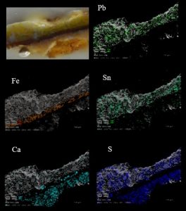
Elemental mapping of SEM-EDS performed on a cross-section of an oil painting. Three pictorial layers are identified, consisting mainly of a surface layer made with lead-tin yellow and white lead (Pb, S, Sn), a medium layer consisting of ocher/earth pigments (Fe), and a preparatory layer of gypsum (Ca, S).
Surface analyses:
The Dafne-Light laboratory is equipped with several portable instruments that allow a non-destructive and non-invasive preliminary analysis of paintings in-situ (archeological sites, museums, restoration laboratories):
Spectroscopic techniques:
- FTIR spectroscopy
- Raman spectroscopy
- UV fluorescence spectroscopy with LED sources (280, 365, 470 nm).
- X-ray fluorescence spectroscopy (XRF)
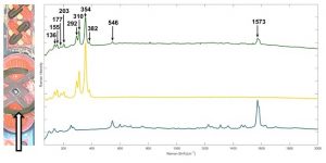
Raman spectroscopy was performed with the portable device on an illuminated manuscript. The green color (green spectrum) has been identified as a mixture of indigo (blue spectrum) and orpiment (yellow spectrum).
Imaging techniques:
- FTIR imaging in reflection mode
- Multispectral imaging (UV-VIS-NIR)
- IR reflectography (900-1500nm)
The identification of modern restorations, repainting and the presence of different varnishes can be evaluated using UV fluorescence photography (the use of sources that block visible radiation permit a more accurate analysis of superficial layers).
Two digital infrared cameras carry out Reflectographs in the near-infrared (750-1000 nm) and in the SWIR (about 1700 nm) with the aim of visualizing underdrawings and other spectral features of the used pigments/dyes.
Finally, a multispectral imaging system (VIS-NIR) permits the “false colors” image analysis but also provides information on the nature of the used pigments and highlights, by a multivariate approach, any spectral features of the pictorial composition not distinguishable by the naked eye.
Photography (right) and SWIR reflectography of an illuminated manuscript (detail). The disappearance of some pictorial layers indicates the use of transparent infrared pigments. Also the black notes fade supposing the use of iron-based inks (i. e. iron-gall ink).

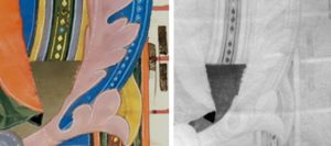
Photography (right) and NIR Reflectography (left) of a fresco. The reflectographic image highlights the restoration additions performed with different pictorial materials than those used for the original painting.
Finally, a multispectral imaging system (VIS-NIR) is used to produce “false colors” image analysis providing information on the nature of the used pigments and highlighting, by a multivariate approach, any spectral features of the pictorial composition not distinguishable by the naked eye.
ARCHEOLOGICAL MATERIALS
In the Dafne-Light laboratory, both organic and inorganic materials coming from archaeological sites can be also analyzed. In addition to the characterization of wall paintings (i. e. frescoes) by using the mentioned techniques in the “pictorial materials” section, it is also possible to perform use-wear analyses and to identify organic residues found on archaeological objects both by means of FT-IR spectroscopy and SEM-EDS.
These two techniques might also provide important information for the study of archaeological bones both to identify the constituent elements and to define the organic and inorganic fractions, as well as their distribution in the case of cross sections.
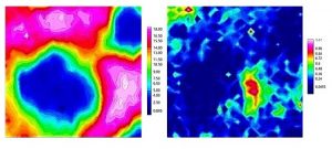
FT-IR chemical mapping of the distribution of collagen (left) and phosphates (right) around osteons of an archaeological bone. The color scale indicates the intensity of the selected absorption bands: pink (MAX value) – blue (MIN value).
MATERIALS FOR RESTORATION AND CONSERVATION TREATMENTS
The identification of some restorative materials such as natural and synthetic resins, waxes, etc … might be performed by using FT-IR spectroscopy both in portable mode and on cross sections. Finally, the Dafne-Light Laboratory deals with the characterization of new-generation materials for the consolidation and conservation of Cultural Heritage by means of FT-IR and Raman spectroscopies (i. e. nano-silica materials).
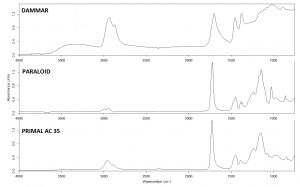
FT-IR (ATR) spectra of a natural resin (dammar paint) and two synthetic varnishes (Paraloid and Primal AC 35).
METALS
The elemental composition of metal samples (smaller than a square centimeter) or cross sections (also embedded in resin) might achieved by means of a scanning electron microscope equipped with microanalysis (SEM-EDS) with the possibility to obtain an elemental distribution map. If the metals are affected by corrosion phenomena, degradation compounds might be detected by means of FT-IR and Raman spectroscopies, which can be performed in situ or on micro-samples.